The Abdomen Sonography Examination (AB-Abdomen)
Passing ARDMS RDMS exam ensures for the successful candidate a powerful array of professional and personal benefits. The first and the foremost benefit comes with a global recognition that validates your knowledge and skills, making possible your entry into any organization of your choice.


AB-Abdomen Exam Dumps
- Exam Code: AB-Abdomen
- Vendor: ARDMS
- Certifications: RDMS
- Exam Name: Abdomen Sonography Examination
Why CertAchieve is Better than Standard AB-Abdomen Dumps
In 2026, ARDMS uses variable topologies. Basic dumps will fail you.
| Quality Standard | Generic Dump Sites | CertAchieve Premium Prep |
|---|---|---|
| Technical Explanation | None (Answer Key Only) | Step-by-Step Expert Rationales |
| Syllabus Coverage | Often Outdated (v1.0) | 2026 Updated (Latest Syllabus) |
| Scenario Mastery | Blind Memorization | Conceptual Logic & Troubleshooting |
| Instructor Access | No Post-Sale Support | 24/7 Professional Help |
Customers Passed Exams
10
Success backed by proven exam prep tools
Questions Came Word for Word
93%
Real exam match rate reported by verified users
Average Score in Real Testing Centre
90%
Consistently high performance across certifications
Study Time Saved With CertAchieve
60%
Efficient prep that reduces study hours significantly
Coverage of Official ARDMS AB-Abdomen Exam Domains
Our curriculum is meticulously mapped to the ARDMS official blueprint.
Liver (22%)
The highest-weighted single-organ domain. Master the segmental anatomy (Couinaud’s), diffuse disease (cirrhosis, fatty infiltration), and focal lesions (hemangiomas, HCC, metastases). In 2026, focus on Elastography basics and the sonographic appearance of Liver Imaging Reporting and Data System (LI-RADS) classifications.
Urinary System (25%)
Deep dive into the kidneys, ureters, and bladder. Master the identification of renal masses, calculi, and vascular abnormalities like Renal Artery Stenosis. Focus on renal transplant evaluation, hydronephrosis staging, and bladder wall pathology.
Gallbladder and Biliary Tree (15%)
Focus on the detection of cholelithiasis, cholecystitis (acute vs. chronic), and biliary obstruction. Master the measurement protocols for the Common Bile Duct (CBD) and the identification of polyps vs. stones using positional changes.
Pancreas (15%)
Mastering the challenging visualization of the pancreas. Focus on pancreatitis (acute vs. chronic), pseudocysts, and neoplastic processes like adenocarcinoma. Understand the relationship between the pancreas and surrounding vasculature (SMA, Splenic Vein).
Abdominal Vasculature (12%)
Focus on the Aorta, IVC, and portal system. Master the criteria for Abdominal Aortic Aneurysms (AAA) and the Doppler hemodynamics of portal hypertension. Learn to identify Budd-Chiari syndrome and Nutcracker syndrome.
Superficial & Miscellaneous Structures (15%)
Includes the Thyroid, Scrotum, and Prostate. Master the ACR TI-RADS for thyroid nodules and the emergent findings in testicular torsion vs. epididymitis. Includes retroperitoneum, abdominal wall, and GI tract (appendicitis/intussusception).
ARDMS AB-Abdomen Exam Domains Q&A
Certified instructors verify every question for 100% accuracy, providing detailed, step-by-step explanations for each.
Question 1
ARDMS AB-Abdomen
QUESTION DESCRIPTION:
Which vessel lies anterior to the uncinate process?
Correct Answer & Rationale:
Answer: D
Explanation:
The superior mesenteric vein (SMV) lies directly anterior to the uncinate process of the pancreas. The uncinate process wraps around the posterior aspect of the SMV and SMA. The portal vein and IVC lie more posteriorly in relation to the pancreatic head.
According to Moore’s Clinically Oriented Anatomy:
“The superior mesenteric vein crosses anterior to the uncinate process of the pancreas.”
[Reference:, Moore KL, Dalley AF, Agur AMR. Clinically Oriented Anatomy. 8th ed. Wolters Kluwer, 2018., Gray’s Anatomy for Students, 4th ed., Elsevier, 2019., —]
Question 2
ARDMS AB-Abdomen
QUESTION DESCRIPTION:
Which condition presents sonographically as an anechoic mass between the umbilicus and the bladder?
Correct Answer & Rationale:
Answer: D
Explanation:
A urachal cyst arises from incomplete closure of the urachus, a remnant of the fetal allantoic duct connecting the bladder to the umbilicus. It appears as a midline, anechoic, nonvascular mass located between the bladder dome and the umbilicus.
According to Rumack’s Diagnostic Ultrasound:
“A urachal cyst is a midline, anechoic structure located between the bladder and umbilicus, resulting from incomplete obliteration of the urachus.”
[Reference:, Rumack CM, Wilson SR, Charboneau JW, Levine D. Diagnostic Ultrasound. 5th ed. Elsevier, 2017., AIUM Practice Parameter for the Performance of Ultrasound of the Pelvis, 2020., —]
Question 3
ARDMS AB-Abdomen
QUESTION DESCRIPTION:
Which description best characterizes a normal systolic spectral waveform of the renal artery?
Correct Answer & Rationale:
Answer: D
Explanation:
A normal renal artery waveform demonstrates rapid systolic upstroke (acceleration) with continuous forward flow in diastole due to the kidney's low-resistance vascular bed. Slow acceleration or blunted peaks may indicate significant renal artery stenosis.
According to Zwiebel’s Introduction to Vascular Ultrasound:
“Normal renal artery waveforms demonstrate a rapid systolic acceleration with a sharp systolic peak.”
[Reference:, Zwiebel WJ, Pellerito JS. Introduction to Vascular Ultrasound. 6th ed. Elsevier, 2019., ACR Practice Parameter for the Performance of a Duplex Doppler Examination, 2021., —]
Question 4
ARDMS AB-Abdomen
QUESTION DESCRIPTION:
Which condition is most likely in a patient presenting with weight loss and fatigue along with elevated liver enzymes, elevated potassium, and decreased sodium?
Correct Answer & Rationale:
Answer: A
Explanation:
Addison disease (primary adrenal insufficiency) results in insufficient production of cortisol and aldosterone. The hallmark laboratory findings include:
Hyponatremia (low sodium)
Hyperkalemia (high potassium)
Elevated liver enzymes (due to nonspecific hepatic involvement)
Fatigue, weight loss, and hypotension are common clinical features.
Conn syndrome (B) causes hyperaldosteronism, leading to hypokalemia (not hyperkalemia).
Acute pancreatitis (C) would typically show elevated amylase/lipase.
Hepatocellular carcinoma (D) may present with elevated liver enzymes but not the electrolyte pattern described.
Reference Extracts:
Nieman LK. "Diagnosis and Treatment of Primary Adrenal Insufficiency." J Clin Endocrinol Metab. 2011;96(7):1957-1966.
Rumack CM, Wilson SR, Charboneau JW, Levine D. Diagnostic Ultrasound. 5th ed. Elsevier, 2017.
—
Question 5
ARDMS AB-Abdomen
QUESTION DESCRIPTION:
Based on this image, which congenital anomaly should be suspected?
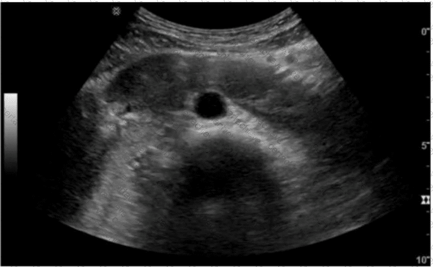
Correct Answer & Rationale:
Answer: C
Explanation:
The ultrasound image demonstrates a dilated duodenum with a hypoechoic soft tissue structure encircling it. This is a classic sonographic appearance suggestive of an annular pancreas. In annular pancreas, pancreatic tissue completely or partially encircles the second portion of the duodenum, which can lead to duodenal narrowing or obstruction.
Annular pancreas is a congenital anomaly that results from failure of the ventral pancreatic bud to rotate properly during embryologic development. As a result, pancreatic tissue encircles the duodenum. It may present in neonates with symptoms of duodenal obstruction or in adults with abdominal pain, pancreatitis, or vomiting.
Ultrasound Findings:
Hypoechoic pancreatic tissue encircling the duodenum
Evidence of duodenal dilatation proximal to the obstruction
“Double bubble” sign may be seen in neonates
Differentiation from other options:
A. Supernumerary kidney: Refers to an accessory kidney. It would be seen in the retroperitoneum and is unrelated to the duodenum or pancreas.
B. Pancreas divisum: A ductal anomaly best diagnosed on MRCP or ERCP. It is not typically visible on conventional ultrasound.
D. Horseshoe kidney: A renal fusion anomaly where the lower poles of the kidneys are fused. It is seen in the pelvis or lower abdomen and does not involve the duodenum or pancreas.
[References:, Rumack CM, Wilson SR, Charboneau JW, Levine D. Diagnostic Ultrasound. 5th Edition. Elsevier, 2018. Chapter: Pancreas, pp. 269–272., Radiopaedia.org. Annular pancreas: https://radiopaedia.org/articles/annular-pancreas, AIUM Practice Parameter for the Performance of Abdominal and Retroperitoneal Ultrasound Examinations, 2020., , ]
Question 6
ARDMS AB-Abdomen
QUESTION DESCRIPTION:
Which vascular condition is most likely associated with the sonographic findings demonstrated in this image?
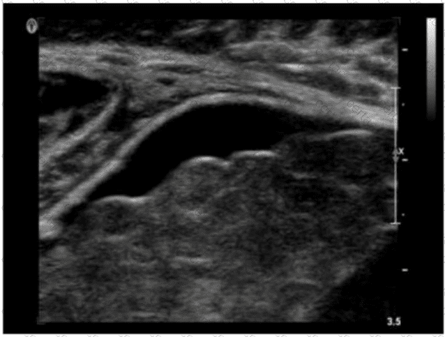
Correct Answer & Rationale:
Answer: C
Explanation:
The ultrasound image demonstrates a tubular, anechoic structure coursing anterior to the left portal vein and heading toward the anterior abdominal wall. This is consistent with a recanalized umbilical vein, which is an important collateral pathway that reopens in cases of portal hypertension.
Normally, the umbilical vein becomes obliterated after birth and forms the ligamentum teres. However, in the setting of significant portal hypertension, the umbilical vein may recanalize and serve as a collateral route to decompress the portal system.
Sonographic features of a recanalized umbilical vein:
Anechoic, tubular structure in the ligamentum teres fissure
Seen anterior to the left portal vein
Color Doppler confirms hepatofugal venous flow
Associated with signs of portal hypertension (e.g., splenomegaly, varices)
Differentiation from other options:
A. Budd-Chiari syndrome: Involves hepatic vein outflow obstruction; ultrasound shows absent or narrowed hepatic veins and may have caudate lobe hypertrophy.
B. Splenic artery aneurysm: Typically visualized near the splenic hilum as a pulsatile cystic mass; Doppler shows arterial flow.
D. Median arcuate ligament syndrome: Involves compression of the celiac axis; best assessed with Doppler showing elevated velocities on expiration.
[References:, Rumack CM, Wilson SR, Charboneau JW, Levine D. Diagnostic Ultrasound. 5th Edition. Elsevier, 2018. Chapter: Portal Hypertension and Collaterals, pp. 101–104., American Institute of Ultrasound in Medicine (AIUM). Practice Parameter for the Performance of a Vascular Ultrasound Examination, 2020., Radiopaedia.org. Recanalized umbilical vein: https://radiopaedia.org/articles/recanalised-umbilical-vein, , ]
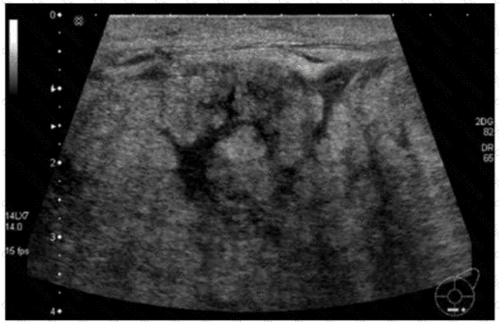
Question 7
ARDMS AB-Abdomen
QUESTION DESCRIPTION:
Which patient maneuver would best aid in identifying the pathology demonstrated in this image?
Correct Answer & Rationale:
Answer: D
Explanation:
The ultrasound image demonstrates a classic example of ascites, shown by the anechoic (dark) fluid located between bowel loops or surrounding abdominal organs. In this case, there appears to be a small fluid collection in the peritoneal cavity.
One of the key maneuvers used to differentiate free fluid (such as ascites) from loculated fluid or other structures is to reposition the patient. Asking the patient to “turn from side to side” (Option D) can help in assessing whether the fluid shifts position — a hallmark feature of free intraperitoneal fluid. This positional change is highly useful in confirming the diagnosis and distinguishing ascites from other potential mimics (e.g., cystic masses, lymphoceles, or bowel wall thickening).
In contrast:
Drinking water (A) is often used in imaging the urinary bladder or gastrointestinal tract but not for fluid characterization.
Standing upright (B) may shift fluid but is less practical during real-time ultrasound.
Breathing quietly (C) doesn’t significantly aid in visualizing peritoneal fluid mobility.
[References:, Rumack CM, Wilson SR, Charboneau JW, Levine D. Diagnostic Ultrasound, 5th ed. Elsevier; 2017., Hagen-Ansert SL. Textbook of Diagnostic Sonography, 8th ed. Elsevier; 2017., AIUM Practice Parameter for the Performance of Diagnostic and Screening Ultrasound Examinations of the Abdomen and/or Retroperitoneum (2020)., , ]
Question 8
ARDMS AB-Abdomen
QUESTION DESCRIPTION:
Which condition is most likely associated with this image of the common bile duct?
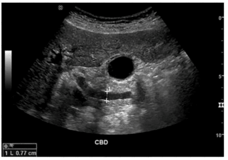
Correct Answer & Rationale:
Answer: C
Explanation:
The ultrasound image demonstrates a dilated common bile duct (CBD), measuring approximately 7.7 mm in diameter. A normal CBD should generally measure less than 6 mm in a patient under 60 years old and may increase approximately 1 mm per decade thereafter or after cholecystectomy.
In the absence of gallstones within the CBD, one of the most concerning causes of CBD dilation is distal obstruction due to an extrinsic compressive lesion. The most common and clinically significant cause of distal CBD obstruction is a mass at the head of the pancreas.
A pancreatic head mass (e.g., adenocarcinoma) may compress the distal CBD and pancreatic duct simultaneously, resulting in the “double duct sign” — dilation of both the CBD and pancreatic duct. This is a classic finding in pancreatic cancer.
Comparison of answer choices:
A. Liver mass — unlikely to cause isolated CBD dilation unless invading the porta hepatis.
B. Cystic duct stone — may cause gallbladder hydrops but typically not CBD dilation unless Mirizzi syndrome is present.
C. Pancreatic head mass — Correct. This is the most likely cause of painless progressive CBD dilation without visible intraductal stones.
D. Gallbladder stones — These may be associated with biliary colic or cholecystitis but typically do not cause CBD dilation unless the stone has migrated and obstructed the distal duct.
[References:, Rumack CM, Wilson SR, Charboneau JW, Levine D. Diagnostic Ultrasound, 5th ed. Elsevier; 2017., Lee JK, Sagel SS, Stanley RJ.Computed Body Tomography with MRI Correlation, 4th ed. Lippincott Williams & Wilkins; 2006., ACR Appropriateness Criteria® Right Upper Quadrant Pain (2021)., , ]
Question 9
ARDMS AB-Abdomen
QUESTION DESCRIPTION:
Which sonographic appearance of the normal epididymis is the most common?
Correct Answer & Rationale:
Answer: C
Explanation:
The normal epididymis typically appears as a homogeneous structure that is either isoechoic or slightly hypoechoic compared to the testis. The most accurate description is "homogeneous compared to the testis," meaning the texture is uniform. It is not anechoic, nor does it typically show irregular borders unless pathology is present.
According to Rumack’s Diagnostic Ultrasound:
"The normal epididymis appears homogeneous and is isoechoic or slightly hypoechoic relative to the testis." (Rumack CM et al., Diagnostic Ultrasound, 5th ed.)
[Reference:, Rumack CM, Wilson SR, Charboneau JW, Levine D. Diagnostic Ultrasound. 5th ed. Elsevier; 2017., AIUM Practice Parameter for Scrotal Ultrasound, 2020., —]
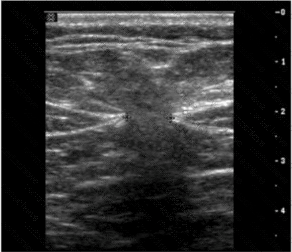
Question 10
ARDMS AB-Abdomen
QUESTION DESCRIPTION:
A lactating female presents with a tender, swollen breast, erythema, and fever. Which condition is most likely present in this image?
Correct Answer & Rationale:
Answer: C
Explanation:
The clinical presentation—tender, swollen breast with erythema and fever—in a lactating female strongly suggests acute mastitis. The sonographic findings support this diagnosis. In the image, the breast parenchyma shows diffuse, hypoechoic, and heterogeneous echotexture with increased vascularity, which is consistent with inflammatory changes typical of mastitis.
Mastitis is a common complication during lactation, particularly in the first few weeks postpartum. It results from milk stasis and subsequent bacterial infection, commonly due to Staphylococcus aureus. Ultrasound features of mastitis include:
Ill-defined, hypoechoic, edematous areas in the breast parenchyma
Increased Doppler flow due to hyperemia
Skin thickening
Ductal dilatation may also be present
If left untreated, mastitis may progress to abscess formation, which would appear as a localized, complex fluid collection with peripheral hyperemia and internal debris. However, the image does not show a well-formed fluid collection consistent with abscess.
Option B (Ductal carcinoma): Inappropriate here due to the acute clinical scenario and patient age. Ductal carcinoma typically presents as a hypoechoic mass with irregular margins and posterior shadowing, not diffuse edema or inflammatory changes.
Option D (Galactocele): This benign milk-filled retention cyst typically appears anechoic or with fluid–fluid levels but lacks signs of inflammation and systemic symptoms such as fever.
Option A (Abscess): This could be a differential, but abscesses usually present with a well-defined anechoic or complex mass. The absence of a discrete collection and the diffuse appearance makes mastitis more likely.
[References:, Mendelson EB. Practical Ultrasound: An Illustrated Guide. Springer, 2004. Chapter: Breast Ultrasound., American College of Radiology (ACR). ACR Practice Parameter for the Performance of a Breast Ultrasound Examination, 2022., Rumack CM, Wilson SR, Charboneau JW, Levine D. Diagnostic Ultrasound. 5th Edition. Elsevier, 2018. Chapter: Breast, pp. 1169–1175., , ]

Verified by Certified Instructors
This ARDMS AB-Abdomen study pack was audited and verified on June 25, 2026 by Eric Huntington,. We ensure every technical rationale aligns with real-world enterprise standards.
A Stepping Stone for Enhanced Career Opportunities
Your profile having RDMS certification significantly enhances your credibility and marketability in all corners of the world. The best part is that your formal recognition pays you in terms of tangible career advancement. It helps you perform your desired job roles accompanied by a substantial increase in your regular income. Beyond the resume, your expertise imparts you confidence to act as a dependable professional to solve real-world business challenges.
Your success in ARDMS AB-Abdomen certification exam makes your visible and relevant in the fast-evolving tech landscape. It proves a lifelong investment in your career that give you not only a competitive advantage over your non-certified peers but also makes you eligible for a further relevant exams in your domain.
What You Need to Ace ARDMS Exam AB-Abdomen
Achieving success in the AB-Abdomen ARDMS exam requires a blending of clear understanding of all the exam topics, practical skills, and practice of the actual format. There's no room for cramming information, memorizing facts or dependence on a few significant exam topics. It means your readiness for exam needs you develop a comprehensive grasp on the syllabus that includes theoretical as well as practical command.
Here is a comprehensive strategy layout to secure peak performance in AB-Abdomen certification exam:
- Develop a rock-solid theoretical clarity of the exam topics
- Begin with easier and more familiar topics of the exam syllabus
- Make sure your command on the fundamental concepts
- Focus your attention to understand why that matters
- Ensure hands-on practice as the exam tests your ability to apply knowledge
- Develop a study routine managing time because it can be a major time-sink if you are slow
- Find out a comprehensive and streamlined study resource for your help
Ensuring Outstanding Results in Exam AB-Abdomen!
In the backdrop of the above prep strategy for AB-Abdomen ARDMS exam, your primary need is to find out a comprehensive study resource. It could otherwise be a daunting task to achieve exam success. The most important factor that must be kep in mind is make sure your reliance on a one particular resource instead of depending on multiple sources. It should be an all-inclusive resource that ensures conceptual explanations, hands-on practical exercises, and realistic assessment tools.
Certachieve: A Reliable All-inclusive Study Resource
Certachieve offers multiple study tools to do thorough and rewarding AB-Abdomen exam prep. Here's an overview of Certachieve's toolkit:
ARDMS AB-Abdomen PDF Study Guide
This premium guide contains a number of ARDMS AB-Abdomen exam questions and answers that give you a full coverage of the exam syllabus in easy language. The information provided efficiently guides the candidate's focus to the most critical topics. The supportive explanations and examples build both the knowledge and the practical confidence of the exam candidates required to confidently pass the exam. The demo of ARDMS AB-Abdomen study guide pdf free download is also available to examine the contents and quality of the study material.
ARDMS AB-Abdomen Practice Exams
Practicing the exam AB-Abdomen questions is one of the essential requirements of your exam preparation. To help you with this important task, Certachieve introduces ARDMS AB-Abdomen Testing Engine to simulate multiple real exam-like tests. They are of enormous value for developing your grasp and understanding your strengths and weaknesses in exam preparation and make up deficiencies in time.
These comprehensive materials are engineered to streamline your preparation process, providing a direct and efficient path to mastering the exam's requirements.
ARDMS AB-Abdomen exam dumps
These realistic dumps include the most significant questions that may be the part of your upcoming exam. Learning AB-Abdomen exam dumps can increase not only your chances of success but can also award you an outstanding score.
Top Exams & Certification Providers
New & Trending
- New Released Exams
- Related Exam
- Hot Vendor