The AE Adult Echocardiography Examination (AE-Adult-Echocardiography)
Passing ARDMS RDCS exam ensures for the successful candidate a powerful array of professional and personal benefits. The first and the foremost benefit comes with a global recognition that validates your knowledge and skills, making possible your entry into any organization of your choice.


AE-Adult-Echocardiography Exam Dumps
- Exam Code: AE-Adult-Echocardiography
- Vendor: ARDMS
- Certifications: RDCS
- Exam Name: AE Adult Echocardiography Examination
Why CertAchieve is Better than Standard AE-Adult-Echocardiography Dumps
In 2026, ARDMS uses variable topologies. Basic dumps will fail you.
| Quality Standard | Generic Dump Sites | CertAchieve Premium Prep |
|---|---|---|
| Technical Explanation | None (Answer Key Only) | Step-by-Step Expert Rationales |
| Syllabus Coverage | Often Outdated (v1.0) | 2026 Updated (Latest Syllabus) |
| Scenario Mastery | Blind Memorization | Conceptual Logic & Troubleshooting |
| Instructor Access | No Post-Sale Support | 24/7 Professional Help |
Customers Passed Exams
10
Success backed by proven exam prep tools
Questions Came Word for Word
91%
Real exam match rate reported by verified users
Average Score in Real Testing Centre
87%
Consistently high performance across certifications
Study Time Saved With CertAchieve
60%
Efficient prep that reduces study hours significantly
Coverage of Official ARDMS AE-Adult-Echocardiography Exam Domains
Our curriculum is meticulously mapped to the ARDMS official blueprint.
Anatomy, Physiology, and Hemodynamics (28%)
Master cardiac anatomy, normal physiology, and complex hemodynamics. Includes pressure gradients, stroke volume calculations, and the continuity equation logic.
Valvular Heart Disease (25%)
Deep dive into Mitral, Aortic, Tricuspid, and Pulmonary valve pathologies. Focus on stenosis, regurgitation, and prosthetic valve assessment using Doppler criteria.
Cardiomyopathies and Other Pathologies (25%)
Comprehensive coverage of Hypertrophic, Dilated, and Restrictive cardiomyopathies, as well as pericardial diseases, systemic hypertension, and cardiac masses.
Acquisition, Protocols, and Measurements (14%)
Mastering TTE and TEE protocols, standard imaging planes, M-mode, and 2D measurements. Focus on ventricular function assessment and strain imaging.
Clinical Safety, Physics, and Instrumentation (8%)
Core knowledge of ultrasound physics, bioeffects, safety protocols, and optimizing instrumentation settings (Gain, TGC, Depth) for adult echo exams.
ARDMS AE-Adult-Echocardiography Exam Domains Q&A
Certified instructors verify every question for 100% accuracy, providing detailed, step-by-step explanations for each.
Question 1
ARDMS AE-Adult-Echocardiography
QUESTION DESCRIPTION:
Which finding is associated with partial anomalous venous return?
Correct Answer & Rationale:
Answer: C
Explanation:
Partial anomalous pulmonary venous return (PAPVR) is a congenital defect where some pulmonary veins drain into the right atrium or systemic venous circulation rather than the left atrium. It is frequently associated with sinus venosus atrial septal defect (ASD), a defect near the junction of the superior vena cava and right atrium.
Cleft mitral valve is commonly associated with atrioventricular septal defects. Persistent left superior vena cava is a separate venous anomaly not typically linked with PAPVR. Perimembranous ventricular septal defects are different congenital defects not related to pulmonary venous anomalies.
The association between PAPVR and sinus venosus ASD is well described in the " Textbook of Clinical Echocardiography, 6e " , Chapter on Congenital Heart Disease and Shunt Lesions 【 20:120-130†Textbook of Clinical Echocardiography 】
Question 2
ARDMS AE-Adult-Echocardiography
QUESTION DESCRIPTION:
When utilizing contrast agents, what should the sonographer keep in mind?
Correct Answer & Rationale:
Answer: A
Explanation:
Contrast agents used in echocardiography can rarely cause anaphylactoid reactions, which are non-IgE-mediated hypersensitivity reactions that can mimic anaphylaxis. Therefore, sonographers must be prepared to manage such reactions.
Contrary to option B, reactions can be severe though rare. Even patients without prior allergies can react. It is incorrect to say the exam poses no risk; proper precautions and monitoring are essential.
These precautions are emphasized in ASE contrast echocardiography guidelines and safety protocols 【 12:ASE Contrast Echocardiography Guidelines†p.190-195 】【 16:Textbook of Clinical Echocardiography, 6e†p.575-580 】 .
Question 3
ARDMS AE-Adult-Echocardiography
QUESTION DESCRIPTION:
Which Doppler signal is used to calculate the pulmonary artery end-diastolic pressure gradient?
Correct Answer & Rationale:
Answer: C
Explanation:
Pulmonary artery end-diastolic pressure (PAEDP) can be estimated noninvasively by measuring the end-diastolic velocity of pulmonary regurgitation (pulmonary insufficiency) using continuous-wave Doppler. The pressure gradient between the pulmonary artery and right ventricle at end-diastole is calculated using the modified Bernoulli equation from this velocity.
Tricuspid insufficiency is used to estimate right ventricular systolic pressure. Tricuspid inflow and pulmonary inflow velocities provide information on diastolic function but not direct pressure gradients.
This method is well validated and included in ASE guidelines for pulmonary hypertension assessment and Doppler hemodynamics 【 16:Textbook of Clinical Echocardiography, 6e†p.300-305 】【 12:ASE Doppler Guidelines†p.110-115 】 .
Question 4
ARDMS AE-Adult-Echocardiography
QUESTION DESCRIPTION:
Which syndrome is associated with pulmonic stenosis?
Correct Answer & Rationale:
Answer: C
Explanation:
Pulmonic stenosis is a congenital valve abnormality often seen in genetic syndromes with cardiac manifestations. Among these, Noonan syndrome is the most frequently associated with pulmonic stenosis. Noonan syndrome is a genetic disorder characterized by distinctive facial features, short stature, and congenital heart defects, with pulmonic valve stenosis being the predominant cardiac lesion. The stenosis is usually valvular and caused by dysplastic pulmonary valve leaflets, leading to obstruction of right ventricular outflow.
Other syndromes listed do not typically present with pulmonic stenosis:
Turner syndrome is more commonly linked with bicuspid aortic valve and coarctation of the aorta, not pulmonic stenosis.
Eisenmenger syndrome refers to the advanced phase of congenital heart defects with significant pulmonary hypertension and is not a genetic syndrome.
Marfan syndrome is predominantly associated with aortic root dilation and mitral valve prolapse, but not with pulmonic stenosis.
This association is well documented in adult echocardiography guidelines and texts, such as the " Textbook of Clinical Echocardiography " by Catherine Otto, which clearly identifies Noonan syndrome as the syndrome most commonly associated with pulmonic stenosis among congenital heart defects 【 16:Chapter on Congenital Heart Disease†Textbook of Clinical Echocardiography, 6e 】 .
Question 5
ARDMS AE-Adult-Echocardiography
QUESTION DESCRIPTION:
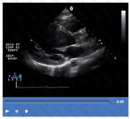
Which pathology is demonstrated in this video clip?
Correct Answer & Rationale:
Answer: D
Explanation:
The video shows prominent trabeculations with deep intertrabecular recesses communicating with the left ventricular cavity, characteristic of isolated left ventricular noncompaction (LVNC). This congenital cardiomyopathy features a spongy myocardial appearance with thickened noncompacted layers.
Amyloidosis typically presents with thickened, bright myocardium but without prominent trabeculations. Sarcoidosis involves granulomatous inflammation, and apical hypertrophic cardiomyopathy shows localized hypertrophy without trabecular changes.
This pathology is detailed in the " Textbook of Clinical Echocardiography, 6e " , Chapter on Cardiomyopathies and Myocardial Disorders 【 20:360-365†Textbook of Clinical Echocardiography 】 .
Question 6
ARDMS AE-Adult-Echocardiography
QUESTION DESCRIPTION:
Which finding does peak mitral valve regurgitant Doppler velocity reflect?
Correct Answer & Rationale:
Answer: D
Explanation:
The peak Doppler velocity of mitral regurgitation (MR) reflects the instantaneous pressure gradient between the left ventricle (LV) and left atrium (LA) during systole. The higher the velocity, the greater the pressure difference.
However, the velocity itself does not quantify severity directly; severity depends on the size and volume of the regurgitant jet. The mechanism is determined by valve morphology and motion, not velocity. The LV to aorta gradient relates to aortic valve pathology.
This principle is discussed in the " Textbook of Clinical Echocardiography, 6e " , Chapter on Mitral Regurgitation and Doppler Evaluation 【 20:390-395†Textbook of Clinical Echocardiography 】 .
Question 7
ARDMS AE-Adult-Echocardiography
QUESTION DESCRIPTION:
What potential source of error is the greatest when calculating the aortic valve area by the continuity equation?
Correct Answer & Rationale:
Answer: C
Explanation:
The continuity equation calculates aortic valve area (AVA) by equating stroke volume through the left ventricular outflow tract (LVOT) to stroke volume through the aortic valve. The equation is:
AVA = (Cross-sectional area of LVOT) × (LVOT VTI) / (Aortic valve VTI)
The cross-sectional area of the LVOT is derived from the LVOT diameter, using the formula π × (diameter/2)^2. Because the diameter is squared in this calculation, even a small error in measurement leads to a significant error in the calculated valve area.
This makes the LVOT diameter measurement the greatest source of error when calculating AVA by the continuity equation. Errors in Doppler velocity measurements (LVOT velocity or aortic jet velocity) are also important but less impactful compared to diameter measurement error.
Aortic valve planimetry is a direct measurement method, not part of the continuity equation. LVOT velocity recorded with pulsed Doppler and aortic jet velocity by continuous wave Doppler are important but not the greatest error source.
This is a well-established concept described in the " Textbook of Clinical Echocardiography, 6e " , Chapter on Valvular Stenosis and Continuity Equation Methodology 【 20:370-375†Textbook of Clinical Echocardiography 】 .
Question 8
ARDMS AE-Adult-Echocardiography
QUESTION DESCRIPTION:
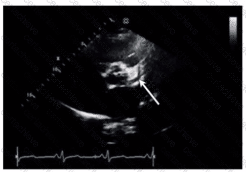
Which coronary artery is identified by the arrow on this image?
Correct Answer & Rationale:
Answer: D
Explanation:
The arrow points to the left anterior descending (LAD) coronary artery, which runs in the anterior interventricular groove toward the apex of the heart. It supplies the anterior wall of the left ventricle.
The right coronary artery runs in the right atrioventricular groove. The left main coronary artery is proximal to the LAD and circumflex arteries. The circumflex artery runs in the left atrioventricular groove posteriorly.
This identification is detailed in the " Textbook of Clinical Echocardiography, 6e " , Chapter on Coronary Artery Anatomy and Echocardiographic Visualization 【 20:150-155†Textbook of Clinical Echocardiography 】 .
Question 9
ARDMS AE-Adult-Echocardiography
QUESTION DESCRIPTION:
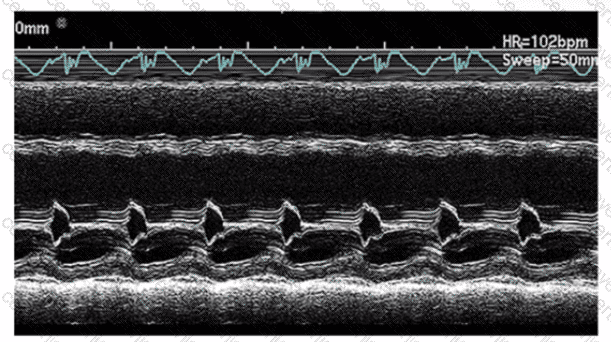
Which condition is most likely demonstrated by this M-mode image?
Correct Answer & Rationale:
Answer: D
Explanation:
The M-mode image shows characteristic diastolic doming or “hockey stick” appearance of the anterior mitral leaflet with restricted leaflet motion. This is a classic sign of mitral stenosis, where leaflet thickening and fusion cause limited opening during diastole.
Dilated cardiomyopathy shows increased chamber sizes and decreased systolic function but not mitral leaflet doming. Hypertrophic cardiomyopathy is characterized by septal thickening and SAM of the mitral valve. Mitral valve prolapse shows leaflet billowing into the left atrium during systole.
This pattern is well described in ASE valvular heart disease guidelines and echocardiography texts 【 12:ASE Valve Imaging Guidelines†p.180-185 】【 16:Textbook of Clinical Echocardiography, 6e†p.200-205 】 .
Question 10
ARDMS AE-Adult-Echocardiography
QUESTION DESCRIPTION:
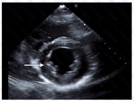
Which wall is indicated by the arrow on this image?
Correct Answer & Rationale:
Answer: B
Explanation:
The echocardiographic image is a parasternal long axis or apical view showing the left ventricle. The arrow points to the wall segment located inferiorly, corresponding to the inferior wall of the left ventricle. The inferior wall is typically visualized in parasternal long axis and apical views as the posterior aspect of the ventricle.
Other options correspond to different walls: anterior is anterior septal wall, anterolateral and inferolateral refer to the lateral wall regions. Accurate wall identification is critical for regional wall motion analysis and coronary artery territory correlation.
This segmental wall identification is detailed in adult echocardiography and ASE chamber quantification guidelines 【 12:ASE Chamber Quantification Guidelines†p.90-95 】【 16:Textbook of Clinical Echocardiography, 6e†p.140-145 】 .

Verified by Certified Instructors
This ARDMS AE-Adult-Echocardiography study pack was audited and verified on June 25, 2026 by Eric Huntington,. We ensure every technical rationale aligns with real-world enterprise standards.
A Stepping Stone for Enhanced Career Opportunities
Your profile having RDCS certification significantly enhances your credibility and marketability in all corners of the world. The best part is that your formal recognition pays you in terms of tangible career advancement. It helps you perform your desired job roles accompanied by a substantial increase in your regular income. Beyond the resume, your expertise imparts you confidence to act as a dependable professional to solve real-world business challenges.
Your success in ARDMS AE-Adult-Echocardiography certification exam makes your visible and relevant in the fast-evolving tech landscape. It proves a lifelong investment in your career that give you not only a competitive advantage over your non-certified peers but also makes you eligible for a further relevant exams in your domain.
What You Need to Ace ARDMS Exam AE-Adult-Echocardiography
Achieving success in the AE-Adult-Echocardiography ARDMS exam requires a blending of clear understanding of all the exam topics, practical skills, and practice of the actual format. There's no room for cramming information, memorizing facts or dependence on a few significant exam topics. It means your readiness for exam needs you develop a comprehensive grasp on the syllabus that includes theoretical as well as practical command.
Here is a comprehensive strategy layout to secure peak performance in AE-Adult-Echocardiography certification exam:
- Develop a rock-solid theoretical clarity of the exam topics
- Begin with easier and more familiar topics of the exam syllabus
- Make sure your command on the fundamental concepts
- Focus your attention to understand why that matters
- Ensure hands-on practice as the exam tests your ability to apply knowledge
- Develop a study routine managing time because it can be a major time-sink if you are slow
- Find out a comprehensive and streamlined study resource for your help
Ensuring Outstanding Results in Exam AE-Adult-Echocardiography!
In the backdrop of the above prep strategy for AE-Adult-Echocardiography ARDMS exam, your primary need is to find out a comprehensive study resource. It could otherwise be a daunting task to achieve exam success. The most important factor that must be kep in mind is make sure your reliance on a one particular resource instead of depending on multiple sources. It should be an all-inclusive resource that ensures conceptual explanations, hands-on practical exercises, and realistic assessment tools.
Certachieve: A Reliable All-inclusive Study Resource
Certachieve offers multiple study tools to do thorough and rewarding AE-Adult-Echocardiography exam prep. Here's an overview of Certachieve's toolkit:
ARDMS AE-Adult-Echocardiography PDF Study Guide
This premium guide contains a number of ARDMS AE-Adult-Echocardiography exam questions and answers that give you a full coverage of the exam syllabus in easy language. The information provided efficiently guides the candidate's focus to the most critical topics. The supportive explanations and examples build both the knowledge and the practical confidence of the exam candidates required to confidently pass the exam. The demo of ARDMS AE-Adult-Echocardiography study guide pdf free download is also available to examine the contents and quality of the study material.
ARDMS AE-Adult-Echocardiography Practice Exams
Practicing the exam AE-Adult-Echocardiography questions is one of the essential requirements of your exam preparation. To help you with this important task, Certachieve introduces ARDMS AE-Adult-Echocardiography Testing Engine to simulate multiple real exam-like tests. They are of enormous value for developing your grasp and understanding your strengths and weaknesses in exam preparation and make up deficiencies in time.
These comprehensive materials are engineered to streamline your preparation process, providing a direct and efficient path to mastering the exam's requirements.
ARDMS AE-Adult-Echocardiography exam dumps
These realistic dumps include the most significant questions that may be the part of your upcoming exam. Learning AE-Adult-Echocardiography exam dumps can increase not only your chances of success but can also award you an outstanding score.
Top Exams & Certification Providers
New & Trending
- New Released Exams
- Related Exam
- Hot Vendor